Avail of diagnostic services from the comfort of your home!
An MRI (Magnetic Resource Imaging) is a scanning procedure that uses a magnetic field, radio waves, and a computer to create precise and thorough images of the human body. The magnetic field’s intensity is measured in teslas or T.
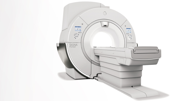
Magnetic resource imaging, as opposed to CT scans, creates 3-dimensional images without the use of dangerous radiation. The magnetic field of an MRI machine causes your body’s hydrogen atoms to momentarily rearrange. These aligned atoms emit very tiny signals when exposed to radio waves, which are utilized to produce cross-sectional MRI pictures that can be stitched together to create 3-D views of various human organs. Fantastic atomic details are produced by the powerful magnetic field produced by the 3.0 Tesla MRI. The photographs produced are sharp and have a lot of resolution. These thorough photographs have improved accuracy and precision in the medical field.
By going down to the ultra-structural level, the 3 Tesla MRI provided at Celera Diagnostics enables the radiologist to see structures that might not be apparent on lower-end equipment, such as blood arteries as small as 200 to 300 microns. The patient spends less time in the machine because of the technology’s ability to perform speedier scanning.
Nearly every part of the body can be seen using an MRI scan at Celara, including lungs, (pulmonary angiogram), blood vessels, bones and joints, spine, abdomen, and pelvis. It is used to:
Routine Scans – Brain, Whole Spine, Abdomen, and Musculoskeletal system
Celara Diagnostics provides MRI, CT scans, ultrasound, X-ray, mammography, lab, cardiology, gastroenterology, and neurology services.
For any Queries or Grievances Contact: Mr. Chandan
Copyright © 2026 Celera Diagnostics,
All rights Reserved.